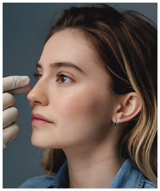
A dorsal hump is the most frequent reason for seeking rhinoplasty. It is seen as a bump on the bridge of the nose in profile; patients find it bothersome in photographs taken from the side, and it can make the face look harsher than it is. On this page I describe what a hump is, how it is removed, and why the request to "just take the hump off" usually calls for more than that.
What is a hump and why does it form?
The bridge of the nose consists of bone in its upper half and cartilage in its lower half. A hump is excess tissue at the junction of these two structures, usually with both a bony and a cartilaginous component. Its causes:
- Heredity: the most common cause. A similar profile is often seen in the family, and the hump becomes apparent with nasal growth in adolescence.
- Trauma: blows in childhood or later can lead to irregular healing and hump formation at the bone-cartilage junction, often with deviation as well.
- A drooping tip: in some patients the hump is not actually large; a drooping tip makes the normal prominence of the bridge look more marked than it is. This distinction changes the plan.
Can only the hump be removed?
This is the question asked most often in consultations. The answer depends on the whole nose. When the hump is removed, the roof of the bridge is opened; a flat, wide area forms between the side bones (an "open roof"). To close it, controlled cuts (osteotomies) are made in the side bones and the roof is brought back together. Otherwise the bridge looks wide and flat from the front. In addition, lowering the bridge changes its relation to the tip: a tip that looked balanced before may look droopy or wide once the hump is gone. For this reason the hump is treated within the whole profile; in some patients a limited tip correction is added.
How is the hump removed?
Several methods are used; the choice depends on the size of the hump, the proportion of bone to cartilage and skin thickness:
- The classical method: the bony part is corrected with a rasp or osteotome, the cartilaginous part with a scalpel. A long-established and predictable method.
- The piezo (ultrasonic) method: the bone is cut and shaped with a fine tip vibrating at ultrasonic frequency; it has less contact with the soft tissues and allows controlled smoothing of bone edges. See the Piezo Rhinoplasty page for details.
- Preservation methods: instead of cutting the hump away, the bridge is kept intact and lowered by removing tissue from beneath it. The natural line of the bridge and the support of the valve area are preserved; however, it is not suitable for every type of hump. Suitability is assessed during examination.
None of these methods is "better" on its own; the right choice depends on the structure of the nose.
Profile planning: how much to remove?
Flattening the hump completely does not look natural on every face. A straight or very slightly curved bridge in women and a straight, strong profile in men are usually perceived as natural; there are also plans in which part of the hump is deliberately kept. An over-resected bridge (a scooped or "ski-slope" look) both looks artificial and is a difficult reason for revision. The amount to be removed is therefore decided to the millimetre and discussed on photographs during the examination. For differences by sex see the Male Rhinoplasty page.
The airway and the hump
The hump sits at the junction of bone and cartilage; this is also where the internal nasal valve is located. If support in this area weakens while the hump is removed, the airway can narrow. As an ENT-trained surgeon I take care in hump surgery to leave the valve area supported or, if necessary, to reinforce it with grafts. If deviation or a septal problem accompanies the hump, it is corrected in the same operation; see Functional Rhinoplasty.
After the operation
Because bone cuts are made in hump surgery, bruising around the eyes may be seen in the first days; swelling subsides largely within the first two weeks. The splint on the nose stays for about a week. The nose needs to be protected from impact and from the pressure of glasses for a few weeks while the bones heal. Small irregularities on the bridge become clearer as swelling subsides; months are needed for the final assessment.
This page is for general information; the method and plan are decided during examination. For other topics see Rhinoplasty: Overview; to arrange an appointment please use the contact page.